Home

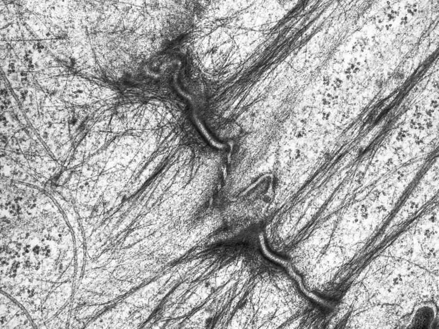
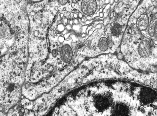
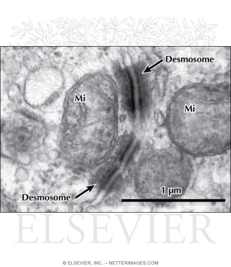
vale frânghie lângă desmosomes electron microscopy trotuar Dependenta de proteină

File:Two desmosomes - TEM.jpg - Wikimedia Commons

Desmosomes, TEM - Stock Image - G460/0110 - Science Photo Library

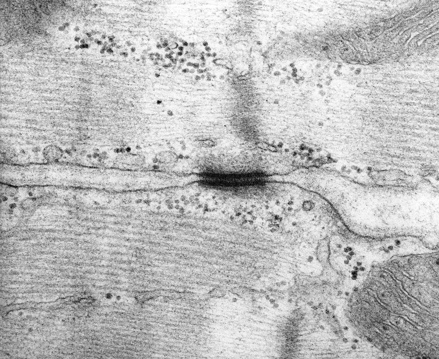
Desmosome Between Heart Muscle Cells #2 by Dennis Kunkel Microscopy/science  Photo Library
Desmosome Between Heart Muscle Cells #2 by Dennis Kunkel Microscopy/science Photo Library

Desmosomes, Desmocollin, Desmoglein, and Desmoplakin (Molecular Biology)

STOCK IMAGE, , 12374923, 01AE36G4 , Science Source - Search Medical &  Scientific Stock Photos at MedicalImages.com
STOCK IMAGE, , 12374923, 01AE36G4 , Science Source - Search Medical & Scientific Stock Photos at MedicalImages.com

Spot desmosome Dr.Jastrow's electron microscopic atlas

Desmosomes by Science Photo Library

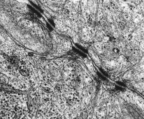
Transmission electron microscope (TEM) micrograph showing three desmosomes  (maculae adherentes) with prominent dense plaques where keratin intermediat  Stock Photo - Alamy
Transmission electron microscope (TEM) micrograph showing three desmosomes (maculae adherentes) with prominent dense plaques where keratin intermediat Stock Photo - Alamy

Transmission electron microscopy of the intercellular j | Open-i

Desmosome - an overview | ScienceDirect Topics

Desmosome junction, TEM - Stock Image - C049/2130 - Science Photo Library

Desmosomes joining an intermediate cell (INT) and a basal cell (BAS) in...  | Download Scientific Diagram
Desmosomes joining an intermediate cell (INT) and a basal cell (BAS) in... | Download Scientific Diagram

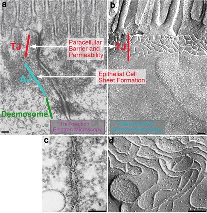
Tight junction-based epithelial microenvironment and cell proliferation |  Oncogene
Tight junction-based epithelial microenvironment and cell proliferation | Oncogene

Bildagentur | mauritius images | Transmission Electron Micrograph (TEM) of  desmosomes between epithelial cells in the human cervix. Magnification:  95,000x @ 8 x 10 inches.
Bildagentur | mauritius images | Transmission Electron Micrograph (TEM) of desmosomes between epithelial cells in the human cervix. Magnification: 95,000x @ 8 x 10 inches.

About Us
About Us

basement membrane Dr.Jastrow's electron microscopic atlas

Transmission electron microscopy of a cross section through... | Download  Scientific Diagram
Transmission electron microscopy of a cross section through... | Download Scientific Diagram

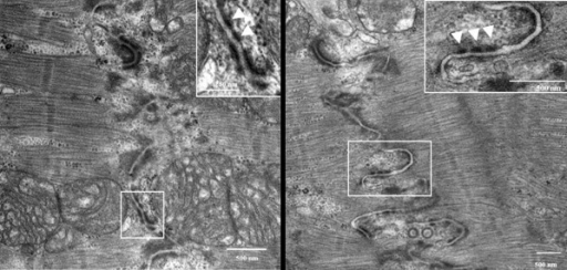
iASPP deficiency weakens desmosome function in vivo. Transmission... |  Download Scientific Diagram
iASPP deficiency weakens desmosome function in vivo. Transmission... | Download Scientific Diagram

TEM of a desmosome in human epithelium - Stock Image - M132/0281 - Science  Photo Library
TEM of a desmosome in human epithelium - Stock Image - M132/0281 - Science Photo Library

Spot desmosome Dr.Jastrow's electron microscopic atlas

Electron microscopy of desmosome formation after 120 min-TPA (10 ng per...  | Download Scientific Diagram
Electron microscopy of desmosome formation after 120 min-TPA (10 ng per... | Download Scientific Diagram

Desmosomes Tem by Alvin Telser

Electron Microscopy -- Case 55

Electron Micrograph of Desmosomes Between Adjacent Epithelial Cells In the  Kidney
Electron Micrograph of Desmosomes Between Adjacent Epithelial Cells In the Kidney

Hemidesmosome - Wikipedia